


New York Times
Eric Li MED ’25 and his three lab partners stared at the coffin-sized metal box. The steel glinted beneath the lights, austere and impenetrable. Through his mask, Li inhaled the faint, familiar smell of formaldehyde.
Only moments before, one hundred of Li’s fellow first-year Yale medical students had filed into the cavernous, glass-paneled laboratory on the third floor of The Anlyan Center for Medical Research and Education, a block away from the Yale School of Medicine. It was their first time in the dissection room. They navigated around rows of narrow steel tables mounted with rectangular metal boxes.
Their instructor, Associate Professor of Surgery William Stewart, announced the assembled students’ first lab assignment. Li and his classmates gripped the handles of the boxes at their lab stations and pulled them open. A semi-transparent plastic bag lay inside. They unzipped it.
A 97-year-old woman faced them. A placard on the wall stated that she had died of cardiopulmonary arrest — caused when the heart and lungs cease to function. Li and his lab partners knew nothing else about the cadaver. For a moment, he struggled to fully grasp what he was seeing.
“[It was] a quite surreal experience,” Li recalled. “That’s somebody who used to live and have a whole life and a whole life story. It felt like we didn’t get that much time to process the fact that this was happening.”
For first-year Yale medical students, dissecting a cadaver is often considered a rite of passage — a necessary practice that elicits excitement and dread. Each year, a new class of aspiring physicians wrestles with the psychological and ethical consequences of this macabre tradition.
Li observed the dead woman’s shriveled, jaundiced skin. It had a waxy sheen. The cadavers, most of whom died in their seventies and eighties, sometimes reminded students of their grandparents. Addy Feibel MED ’25, a Yale medical student in Li’s class, had witnessed bodies at open-casket funerals and was mostly unfazed, though she had never been in the presence of twenty-six cadavers at once.
Under Stewart’s instruction, Li and his classmates drew a part of their donor’s body, the body they would study for the next year and a half. They sketched sharp profiles, contoured hands, clusters of freckles and tattoos. Then, the students used marking pens to trace the areas where they imagined they’d find each organ.
They did not actually dissect the body until their third class. They made their first incision with scalpels, slicing through the torso to access the body cavity, navigating dense layers of striated muscle and dull, tan subcutaneous fat. The intestines and lungs glistened beneath the lights. The blood vessels were clouded, with thick clots that, when extracted, formed delicate networks like tree branches. They were nothing like the vibrant vessels illustrated in the medical textbooks Li pored over in his classes. Unlike a live body with richly colored, wet tissue, the donor’s abdomen was mostly sapped of moisture. Yet fluids gushed into the body cavity once Li removed the lungs and set them on the table.
———
The Desensitization of First-Year Medical Students
Over the course of twenty labs spanning a year and a half, Li and his classmates dissected every part of the donor, from the orbital muscles to the gluteus maximus, as generations of doctors had done before them. But the procedures took a toll. “A lot of the times I felt it easiest for me if I went on an autopilot mode and just went about the dissection as a learning exercise or as an almost objective kind of thing. Like I’m going in here to look for these structures, and then I am done,” Li said.
Given the invasiveness of the procedures they perform, Feibel and Li said that the desensitization of medical students is inevitable. It becomes a necessary tool for survival, they said.
Feibel, for instance, recalled feeling hungry every time she emerged from the anatomy laboratory. (Formaldehyde is well-known among medical students as an appetite stimulant). At first she found it unsettling that she craved food after hours of dissecting necrotic tissue. But eventually, “it becomes normal, not weird anymore, to be hungry afterwards,” Feibel said. “It’s just the inside of a body. It’s kind of beautiful. [You] have to get used to what could be perceived by other people as kind of gross because that’s what you have to do when you’re doing surgery.
The dissection of the head and neck, however, punctured the students’ protective emotional boundary. Most of the time, they kept the face of the cadaver covered. But on the day that they extracted the brain, Li confronted the donor’s face. For the next few hours, the room was filled with the ear-splitting sounds of electric saws cutting through twenty-six skulls, cleaving the center of the face and exposing the maxillary sinuses. Bone particles floated in the air. They wore masks to prevent themselves from inhaling the toxic dust.
After drilling through the skull, Li and his lab partners removed the brain. They extracted a yellow sheath of nerves that trailed from the brain to the floor: the spinal cord. Li held the cadaver’s central nervous system—the source of a lifetime of sensations, thoughts, and feelings — in his hands.
“A lot of the general population has not seen what the underside of a brain looks like in person. Not in a picture, but right there, live, in front of you,” Li said. “That’s stuff that you either desensitize yourself towards, or you don’t, and you always feel that sense of unease.”
Dissecting a once-living human cadaver introduces a host of ethical issues. How does one treat the donor with respect and dignity while conducting a violent, invasive procedure? And how does one reckon with the knowledge that the donor’s body may have been obtained in an unethical manner?
While they continue to struggle with the first question, Li and Feibel were not forced to grapple with the second because the process of procuring the bodies is highly regulated. According to Stewart, each individual donates their body directly to the Yale School of Medicine. The donors are embalmed in the Anlyan Center. After the dissection, the bodies are cremated and sent back to their families, or to the Evergreen Cemetery, two miles west of campus. Medical students are not involved in the process.
“I’m just a cog in the system,” Li said. “The donors voluntarily gave up their bodies; I’m at school; the school got these [cadavers]; I’m in a class; they put me here. I’m not going out, robbing graves myself and dissecting. I know that’s very black and white, but I’m not constantly having moral dilemmas while dissecting.”
———
The Birth of Anatomical Dissection in the United States
Anatomical dissections were incorporated into Western medical education in 1231, when Holy Roman Emperor Frederick III decreed that universities training physicians must hold a public dissection once every five years. The practice was seen as a way for medical students to probe into the depths of the body to gain empirical knowledge of its inner structures.
Dissection emerged as a practice in the United States six centuries later, at a time when orthodox physicians (usually European American, upper middle-class men) were struggling to build their reputations. Denounced as murderers by rival botanic and homeopathic healers due to their aggressive therapies, orthodox physicians relied on dissections as a way to legitimize their practice. They established state medical societies and medical schools outfitted with anatomy labs that excluded alternative healers. One such institution was the Yale School of Medicine, erected in 1810 across from the Grove Street Cemetery.
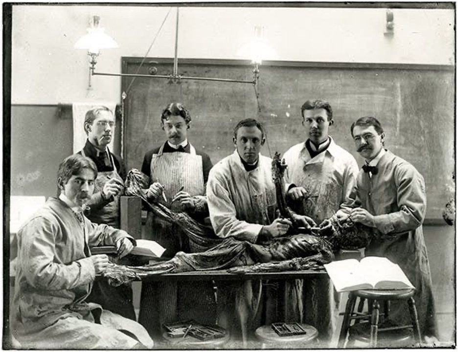
As hundreds of for-profit medical colleges sprung up around the country, orthodox medical students began to identify themselves as anatomists. According to John Warner, the Avalon Professor of the History of Medicine at Yale, the invention of cheap, accessible Brownie cameras in the 1890s popularized group portraits of medical students standing beside their cadavers in the anatomy lab. These photographs became an enduring symbol of orthodox doctors’ professional identity.
“[Posing in the anatomy lab] was the most common way that American medical students, for about a 50-year period, chose to have themselves depicted both together and at work,” Warner said.
Portraits from the late 1890s through the 1920s, unearthed from the archives of the Cushing/Whitney Medical Library, show Yale medical students looking up at the camera mid-dissection, wielding glinting scalpels in their bare hands. Pipes, intended to dispel the odor, dangle from their mouths. The cadaver rests in front of them on the table, fully exposed and rendered unrecognizable by the dissection. Oftentimes, anatomy tables during this period were engraved with racist epigraphs or, more commonly, the sentiment: “He lived for others. He died for us.”
In the late nineteenth and early twentieth centuries, dissection was frequently an act of racist or classist violence. Medical colleges were in constant need of cadavers since there were few legal sources of bodies. Students ventured into nearby graveyards with shovels to “resurrect” the cemetery’s most vulnerable citizens, typically poor African Americans. The dissectors were mostly upper-middle class white men.
Grave robbing was common in New Haven. In 1824, for instance, the body of Bathsheba Smith, a nineteen-yearold white woman, was stolen from her grave in West Haven. Her body was found by the townspeople on the dirtpacked cellar floor of the Yale Medical School, hastily covered in grave clothes. The Connecticut Herald covered the story on January 13, 1824, and the body-snatching sparked a riot on the New Haven Green.
According to Warner, about ten years later, in the 1830s, a similar, little-known incident occurred: a Black man was exhumed from his grave and carried through the streets, witnessed by a crowd of New Haven residents.
“In this case, there’s not a word about it in the New Haven paper [from that time period],” Warner said. “It’s a theft of a black man’s body, and there’s just silence. And that contrast, I think, speaks a lot.”
Today, disproportionately few Black people are willing to donate their bodies to science. According to a study conducted at the University of Massachusetts Memorial Medical Center (UMMMC) in 2018, the overwhelming majority of UMMMC’s 859 donors were white, outnumbering donors of color 82.4 to 17.6 percent and African Americans 82.4 to 4.9 percent.
Over the past few years, however, Stewart has seen the number of Black donors gradually rise in the Yale anatomy program—suggesting renewed trust among communities of color in the medical education system.
———
Haunted by the Past: Yale’s Present Relationship with Anatomical Dissection
He lived for others. He died for us. The black-and-white anatomical portraits preserved in the Cushing/Whitney Medical Library seem like relics of a darker, crueler past far removed from the sterile world of modern medicine. Yet this legacy has reverberated through the decades.
In June 2017, two graduate dental school students and a University of Connecticut orthodontics professor snapped a selfie beside two severed heads intended for research during a workshop held at the Yale Medical School.
Two years later, the Yale Daily News reported that a collection of Yale School of Medicine yearbooks published in the 1990s and in 2011 contained photographs of students dissecting uncovered cadavers, often accompanied by inappropriate captions. In one photograph, a student posed beside the detached arm of a cadaver with the caption, “Hand job.” In another, a group of five medical students stood behind a donor whose exposed face was penetrated by a surgical tool jutting diagonally from their forehead, evoking a murder scene. The words below read: “[A medical student depicted in the photograph] is thinking, ‘One down, four to go.’” In a third, four students and an instructor clustered around a cadaver held up a banner with the words: “She died so that we may learn.” The caption in the 1999 yearbook stated: “’Nuff said.” The photo was eerily similar to portraits of medical students in the 1910s.
Stewart said that while students often resorted to humor to relieve the discomfort associated with dissection, he had never witnessed any student blatantly disrespect the donors.
“I wasn’t very happy [to see those photographs]. It did seem pretty disrespectful to me,” Stewart said.
Warner similarly expressed his dismay with the yearbook photographs. “It’s a lapse of judgment. It is a lapse of professionalism. This is very much the kind of thing that we hope to give a different forum for talking about, reflecting on, and feeling what you’re feeling, but being responsible about how you manage that,” he said.
After the publication of the News article in 2019, Stewart enforced a strict “no photography” policy in the laboratory, which had been introduced ten years before but was previously much more lenient. Now, students sign a code of conduct requiring that they behave respectfully and not disclose details of the lab with others. Before they enter the anatomy lab, Warner also provides each class of medical students with context about the history of dissection and opens a space for them to talk about their affective responses.
Warner acknowledged that dark humor has long been “infused into the fabric of dissecting culture.” Feibel expressed that while she and her labmates dissected their cadaver, they often made jokes, admitting that “the environment is a lot more light-hearted than you [would] think.” Yet she and her friends ensured that they always treated the donors with respect.
“These people have donated their bodies to us, which is a huge honor,” Feibel said. “I think it’s important that we respect them just like we would any patient and not talk about them that way. They’re kind of [like] our first patient.”
Because students work on the same cadaver for a year and a half, Stewart said they often develop a spiritual connection with their donor. At the end of the course, the School of Medicine hosts a Service of Gratitude, in which students present original poetry and artwork dedicated to their donors. The sentiment, Stewart said, is one of overwhelming appreciation. Student artwork hangs in the corridor outside of the lab: a patchwork quilt of a peony sewn by the YSM class of 2005; a knitted set of lungs, intestines, and kidneys from 2008; gorgeously rendered sketches of outstretched hands.
———
Vital Tradition or Dying Practice: The Future of Anatomical Dissection
The cultural and social implications of anatomical dissections are complex. Given their harrowing history, some medical schools have begun to question the necessity of anatomy labs. In 2018, the Cleveland Clinic Lerner College of Medicine announced its revamped “cadaver-less” anatomy program, touted as a new innovation in medical education. The Cleveland school relies on three-dimensional renderings of bodies in virtual reality, coupled with CT scans and ultrasound images of real patients, to teach students anatomy. Other institutions have since followed suit, eliminating anatomy labs from their curricula. The Yale School of Medicine has kept its lab. Yet both Feibel and Li believe that anatomical dissection, while potentially helpful to students, is unnecessary.
Beyond the ethical implications, Li, who wants to specialize in ophthalmology, conveyed that anatomy lab doesn’t have direct clinical applications, even for aspiring surgeons.
“It’s a very controlled environment,” Li said. “You have instructors telling you what structures are. I think if you were actually in an [operating room] seeing a procedure, a real, live, breathing, blood-pumping body looks completely different from a cadaver. Is it beneficial to dissect and learn via this method? Sure. Is it necessary? Not really.”
For Stewart, however, who has taught anatomy at Yale for the past 44 years, anatomical dissection is an irreplaceable part of the medical curriculum. He falls into the empiricist camp, believing that learning occurs not when the student “sees” something, but when they search for and “find” it.
“When you’re touching [the donor], and you’re emotional, and you’re being encouraged by your friends, and you smell it, all of these contribute to the learning. So if you take away, one by one, all of those other senses, you’re left with, in my view, a pale imitation of the actual learning experience,” Stewart said.
Anatomical dissections were once a symbol of the elite status of physicians: pioneers on the frontier of science, excavating the hidden mysteries of the body. Now, some view them as representations of the ethical failures of the medical profession. Regardless of whether dissections fall into obsolescence or remain a time-honored element of modern medical education, they have enabled over 212 generations of Yale medical students to experience the wonders of the human body in ways few will ever appreciate.
“Most people have the idea that dissection is a rite of passage: it’s something that everybody has to go through, [and] it’s miserable,” Stewart said. “We surely don’t want it to be like that. At Yale, we want it to be an experience where people enjoy the camaraderie of the people they’re working with, they enjoy the beauty of the body, they have an insight into how it works, and if it makes them feel better, they [forge] a spiritual connection [with] the donor. We hope that transfers to their patients as well.”